Chest X-Ray
A chest x-ray is a procedure used to evaluate organs and structures within the chest for symptoms of disease. Chest x-rays include views of the lungs, heart, small portions of the gastrointestinal tract, thyroid gland and the bones of the chest area.
Chest x-rays may be performed in a physician’s office, an outpatient radiology facility or hospital radiology department. In some cases, particularly for bedridden patients, a portable chest x-ray may be taken. Portable films are sometimes of poorer quality than those taken with permanent equipment, but are the best choice for certain situations.

Lead plasterboard
Forms a complete shielded envelope to the room. The shielding usually extends to the full structural height of the room.

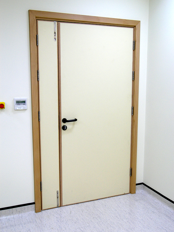
1.5 Lead lined door
A 1.5 leaf door set is usually used for patient entrance. This allows the passage of the patient trolley.

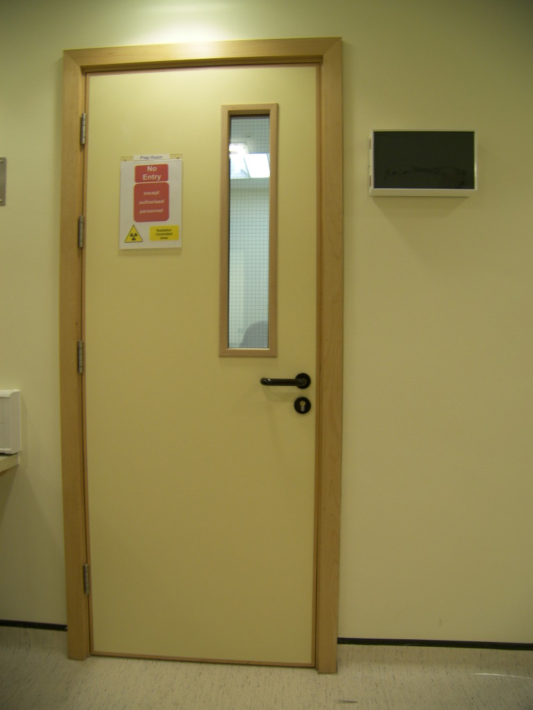
Single door set
A single door set is required as passage from the scanner room to the control room.

Viewing window
Shielded frame and lead glass window enables the operator to see procedures from the control room.